Home
/ Diagram Of The Muscles In The Forearm - Muscles Of The Forearm _ It rotates the forearm and also flexes the elbow.
Diagram Of The Muscles In The Forearm - Muscles Of The Forearm _ It rotates the forearm and also flexes the elbow.
Diagram Of The Muscles In The Forearm - Muscles Of The Forearm _ It rotates the forearm and also flexes the elbow.. 2, ulna, 3, biceps muscle; Yoga anatomy anatomy study anatomy reference anatomy drawing hand therapy massage therapy physical therapy occupational therapy muscle diagram. Your subclavius muscle, which is very often the cause of muscle pain in the upper arm, is located here. The pronator teres and quadratus control pronation, or rotation of the forearm so that the palm faces downward. Commonly known as the bicep muscle, this muscle rests on top of the humerus bone.
Attached to the bones of the skeletal system are about 700 named muscle. It rotates the forearm and also flexes the elbow. Deep muscles, which lie near to the bones (radius and ulnar) or internal organs; Learn vocabulary, terms, and more with flashcards, games, and other study tools. Yoga anatomy anatomy study anatomy reference anatomy drawing hand therapy massage therapy physical therapy occupational therapy muscle diagram.
Muscle Compartments Of The Forearm Complete Anatomy from s3-us-west-1.amazonaws.com Superficial muscles, which lie close to the skin; Photo of arm muscle model with outlined and named muscles. Yoga anatomy anatomy study anatomy reference anatomy drawing hand therapy massage therapy physical therapy occupational therapy muscle diagram. Muscles, for example, exert far greater forces than we might think. It is called lister's tubercle. Begin by massaging the area just below your collarbone. The forearm muscles in the anterior compartment flex the forearm, wrist, and fingers. Figure 1 shows a forearm holding a book and a schematic diagram of an analogous lever system.
Superficial muscles, which lie close to the skin;
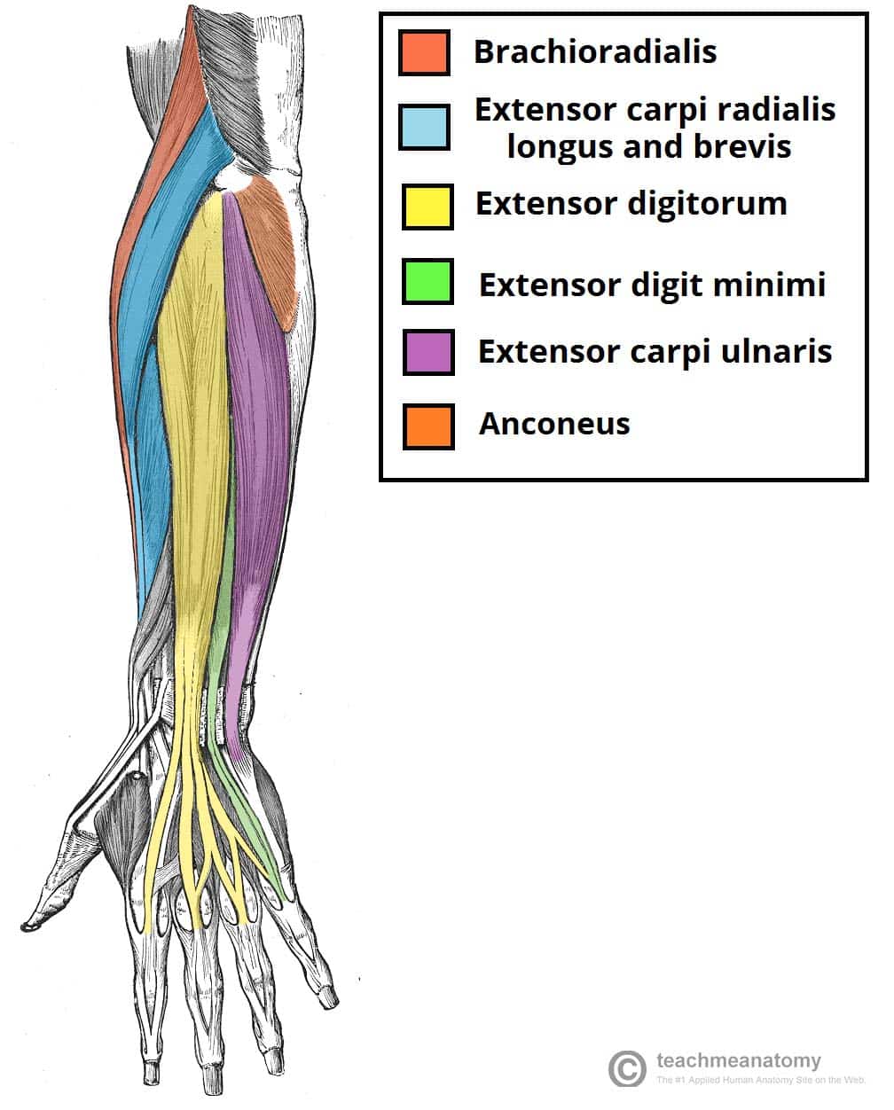
5 minutes the radial musculature consists of three muscles located at the lateral forearm.they all run from or near the lateral epicondyle of the humerus to the wrist. May 31, 2021 · extensor carpi radialis longus muscle (musculus extensor carpi radialis longus. Its muscle belly is in the forearm and the tendon travels along the wrist and enters the third compartment of the band that holds the tendons in position at the wrist. The extensors, which bend lie on the outer side of the forearm and bend it back. Here you can see all the extensor forearm muscles clearly labeled. To begin, spend some time looking at the forearm muscles diagram above. Deep fascia of the forearm).—the antibrachial fascia continuous above with the brachial fascia, is a dense, membranous investment, which forms a general sheath for the muscles in this region; Forearm flexion forearm flexion is rotation in the anatomic plane. Smartdraw includes 1000s of professional healthcare and anatomy chart templates that you can modify and make your own. Similar to the upper arm, the forearm contains an anterior and posterior compartment. This muscle helps rotate the upper arm. The forearm muscles in the anterior compartment flex the forearm, wrist, and fingers. Biceps are large muscle of the upper arm is formally known as the biceps brachii muscle, and rests on top of the humerus bone.
Photo of arm muscle model with outlined and named muscles. Yoga anatomy anatomy study anatomy reference anatomy drawing hand therapy massage therapy physical therapy occupational therapy muscle diagram. Muscles of the arm and forearm diagram, human muscles, muscles of the arm and forearm diagram. Start studying muscles of forearm and wrist 2. / the antibrachial or forearm muscles may be divided into a volar and a dorsal group.
Arm Muscles Anatomy Function Of Biceps Triceps Forearms Openfit from cdn.prod.openfit.com The forearm muscles in the anterior compartment flex the forearm, wrist, and fingers. This forearm muscle is responsible for extending. Muscles of the arm and forearm diagram, human muscles, muscles of the arm and forearm diagram. All muscles in this layer originate from the medial epicondyle of the humerus, they are the flexor carpi ulnaris, flexor carpi radialis, pronator teres and palmaris longus. / the antibrachial or forearm muscles may be divided into a volar and a dorsal group. This bone runs down from the shoulder socket and joins the radius and ulna at the elbow. Photo of arm muscle model with outlined and named muscles. Nov 17, 2014 · the large bones of the arm include:
Photo of arm muscle model with outlined and named muscles.
May 31, 2021 · extensor carpi radialis longus muscle (musculus extensor carpi radialis longus. What is flexor carpi ulnaris? Movements of the wrist, hand, and. It is a functionally important muscle that contains two heads. The forearm muscles in the anterior compartment flex the forearm, wrist, and fingers. Yoga anatomy anatomy study anatomy reference anatomy drawing hand therapy massage therapy physical therapy occupational therapy muscle diagram. The biceps brachii flex the forearm and work with the supinator of the forearm to rotate it so the palm faces upward. The pronator teres and quadratus control pronation, or rotation of the forearm so that the palm faces downward. 2.2 muscle pain in the upper arm: Biceps are large muscle of the upper arm is formally known as the biceps brachii muscle, and rests on top of the humerus bone. This forearm muscle is responsible for extending. Like the upper arm muscles, the forearm muscles can be divided into two parts: Superficial muscles, which lie close to the skin;
Biceps are large muscle of the upper arm is formally known as the biceps brachii muscle, and rests on top of the humerus bone. Figure 1 shows a forearm holding a book and a schematic diagram of an analogous lever system. The biceps brachii flex the forearm and work with the supinator of the forearm to rotate it so the palm faces upward. Diagram of the forearm extensors superficial extensors consist of seven muscles; Movements of the wrist, hand, and.
Muscles Of The Posterior Forearm Superficial Deep Teachmeanatomy from teachmeanatomy.info Your subclavius muscle, which is very often the cause of muscle pain in the upper arm, is located here. What is the superficial layer of anterior muscles of the forearm? Flexors & extensors of the forearm. Figure 1 shows a forearm holding a book and a schematic diagram of an analogous lever system. It is called lister's tubercle. / the antibrachial or forearm muscles may be divided into a volar and a dorsal group. Working with the flexor pollicis longus of the forearm, the flexor pollicis brevis flexes the thumb to grip objects or make a fist. This muscle helps rotate the upper arm.
The forearm is the lower part of the arm, from the elbow to the wrist.
The extensors, which bend lie on the outer side of the forearm and bend it back. Photo of arm muscle model with outlined and named muscles. Anatomy organs human body anatomy human anatomy and physiology forearm muscle anatomy forearm muscles muscle diagram body diagram interactive anatomy upper limb anatomy. Working with the flexor pollicis longus of the forearm, the flexor pollicis brevis flexes the thumb to grip objects or make a fist. These muscles work together to provide a wide range of motion to the little finger. It is called lister's tubercle. Forearm flexion forearm flexion is rotation in the anatomic plane. This bone runs down from the shoulder socket and joins the radius and ulna at the elbow. Nov 17, 2014 · the large bones of the arm include: Yoga anatomy anatomy study anatomy reference anatomy drawing hand therapy massage therapy physical therapy occupational therapy muscle diagram. May 31, 2021 · extensor carpi radialis longus muscle (musculus extensor carpi radialis longus. They also perform pronation, which is to say turning the palm down .they're divided into three layers; And intermediate muscles, which lie between the superficial and deep.